

The arm, or "upper arm" in common usage, is the region of the upper limb that extends from the shoulder to the elbow joint Elbow joint The elbow is the synovial hinge joint between the humerus in the upper arm and the radius and ulna in the forearm. The elbow consists of 3 joints, which form a functional unit enclosed within a single articular capsule. The elbow is the link between the powerful motions of the shoulder and the intricate fine-motor function of the hand. Elbow Joint: Anatomy and connects inferiorly to the forearm Forearm The forearm is the region of the upper limb between the elbow and the wrist. The term "forearm" is used in anatomy to distinguish this area from the arm, a term that is commonly used to describe the entire upper limb. The forearm consists of 2 long bones (the radius and the ulna), the interosseous membrane, and multiple arteries, nerves, and muscles. Forearm: Anatomy through the cubital fossa Cubital Fossa The cubital fossa is the region anterior to the elbow joint. The cubital fossa is seen as the triangular depression between the brachioradialis and pronator teres muscles. The 4 important structures of the cubital fossa (from lateral to medial) are the radial nerve, tendon of the biceps brachii muscle, brachial artery, and median nerve. Cubital Fossa: Anatomy. It is divided into 2 fascial compartments (anterior and posterior) and houses numerous structures, including the humerus; the brachial and profunda brachii arteries Arteries Arteries are tubular collections of cells that transport oxygenated blood and nutrients from the heart to the tissues of the body. The blood passes through the arteries in order of decreasing luminal diameter, starting in the largest artery (the aorta) and ending in the small arterioles. Arteries are classified into 3 types: large elastic arteries, medium muscular arteries, and small arteries and arterioles. Arteries: Histology; the basilic, cephalic, and brachial veins Veins Veins are tubular collections of cells, which transport deoxygenated blood and waste from the capillary beds back to the heart. Veins are classified into 3 types: small veins/venules, medium veins, and large veins. Each type contains 3 primary layers: tunica intima, tunica media, and tunica adventitia. Veins: Histology; the radial, median, ulnar, and musculocutaneous nerves; and the flexor and extensor muscles of the arm.
Last updated: Aug 13, 2026
Contents
Cephalosporins Cephalosporins Cephalosporins are a group of bactericidal beta-lactam antibiotics (similar to penicillins) that exert their effects by preventing bacteria from producing their cell walls, ultimately leading to cell death. Cephalosporins are categorized by generation and all drug names begin with “cef-” or “ceph-.” Cephalosporins are members of the beta-lactam Beta-Lactam Penicillins drug family and consist of:
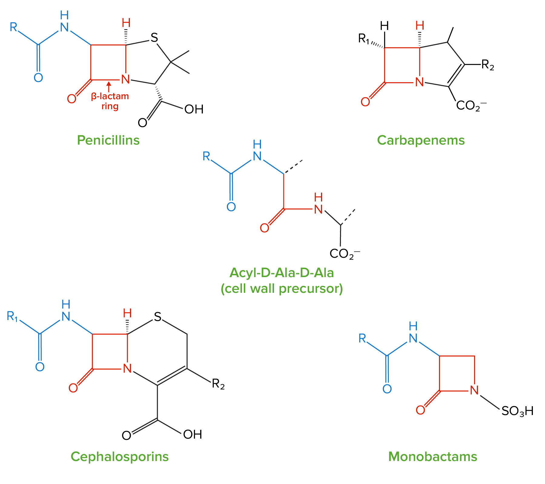
Structure of beta-lactams:
All beta-lactam antibiotics contain the same core 4-membered “beta-lactam” ring (highlighted in red). This ring is responsible for the antibacterial properties of the drug because it is the region that binds to and inhibits the penicillin-binding proteins (PBPs). The PBPs catalyze the formation of the cell wall by generating crosslinks between the peptide chains in the peptidoglycan molecules; PBPs form these crosslinks between acyl-D-Ala-D-Ala peptides, which have a structure similar to the beta-lactam ring.